Isi Email Address Disini:

Kamis, 24 Januari 2013

Mesothelioma Stages

How is mesothelioma staged?
Mesothelioma StagesMesothelioma stages are the assessment levels that doctors utilize to determine the extent of the cancer within the body. Mesothelioma is staged between 1 and 4, depending on severity. Stage 1 mesothelioma, for instance, is highly localized disease, with the tumor affecting only a limited area and organ tissue. Stage 4 cancer, on the other extreme, is extensive disease, which has spread far beyond the tumor’s origin, affecting other organ tissue and even blood or bone cells.
Given how rare mesothelioma is, a formal staging classification exists only for pleural mesothelioma, which affects the lining of the lung and chest cavity. Staging is determined at diagnosis, using any number of diagnostic procedures. Basic staging can be determined through the use of imaging scans, which will provide cancer specialists a visual representation of the extent of the tumor within the body. If the results of imaging scans are inconclusive or it appears that the disease is not localized, a physician may request the patient to undergo a needle or surgical biopsy, which can determine if the malignant cells have metastasized to the blood or lymph nodes through laboratory testing.

Mesothelioma Staging Systems

Today, there are three primary staging systems used to assess how far mesothelioma cancer has spread and each system uses four stages to describe the progression of the disease. It is the definition of each stage within the various systems that can vary slightly.

Butchart Staging System

The Butchart staging system is the oldest and most commonly utilized staging system for mesothelioma. This system is focused on defining the location of the primary tumor mass in the body for each stage. The system doesn’t address how many cancer cells are present, how big the tumor is or the level of cancer present in the body overall.

TNM Staging System

The TNM Staging System, developed by the American Joint Committee on Cancer (AJCC), is similar to staging systems used for other types of cancer. It considers the characteristics of the tumor (T), whether or not lymph nodes are involved (N) and if the cancer has metastasized to other locations in the body (M).

Brigham Staging System

The Brigham Staging System also has four stages of progression and is very similar to the TNM Staging System. The primary difference between the two is that in addition to defining the location of the tumor, and assessing lymph node involvement and the presence of metastatic disease, the Brigham System also helps assess the possibility for and effectiveness of surgical intervention at each stage.

The Stages of Mesothelioma

The four stages of mesothelioma, as noted above, vary slightly within each system but can be generally characterized as indicated below. Please click into the individual pages to get more information about each stage in general and as is it defined within each staging system:

Stage 1 Mesothelioma

In Stage 1 the tumor is localized, there is no lymph node involvement and the cancer has not spread to other organs or tissues. In this case, the cancer is likely restricted to one side of the pleura and surgical removal is possible

Stage 2 Mesothelioma

In Stage 2, the tumor is larger and has invaded the lung or diaphragm. Lymph nodes may also be involved. In this case, surgical resection may be possible though the cancer has likely spread to both sides of the pleura.

Stage 3 Mesothelioma

In Stage 3, mesothelioma has invaded a single region or area such as the chest wall, esophagus, lymph nodes and surgical resection is generally ruled out as an effective treatment.

Stage 4 Mesothelioma

In Stage 4, mesothelioma has invaded multiple regions such as different areas of the chest wall, the diaphragm and/or the pericardium. Lymph glands are also involved and the cancer has spread to other organs. Surgical removal provides no value in this case as the disease has likely metastasized well beyond its origin.

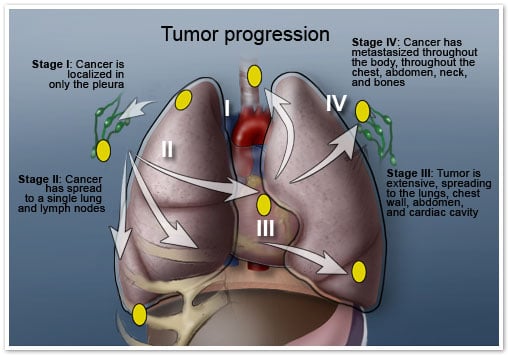
Read more: http://www.mesothelioma.com/mesothelioma/stages/#ixzz2ItmZ7t8d
Read More...

Mesothelioma Biopsies

Mesothelioma BiopsiesPatients that are exhibiting mesothelioma symptoms such as a large buildup of fluid in the chest or abdomen, and that have a history of asbestos exposure, will frequently be sent for a biopsy of the fluid and/or tissue by their doctor. There are two primary types of biopsies that mesothelioma patients may undergo: needle biopsies and surgical biopsies.
Mesothelioma is a disease that is characterized by a long latency period and symptoms that often mimic those of more minor upper respiratory conditions. As a result, diagnosis of this cancer is often delayed. Frequently, by the time that a patient is diagnosed with the disease, it has progressed to advanced stages where therapeutic treatment options become more limited.
This section discusses the types of biopsies that may be performed, which biopsy procedures are recommended depending on where the fluid buildup is occurring and the types of information that can be acquired through biopsy to aid in the diagnosis and treatment of mesothelioma.

Needle Biospy

A fine needle biopsy is considered to be a closed biopsy procedure and therefore less invasive than open surgical biopsies. Pleural biopsy is the most common closed biopsy procedure that is performed to aid in the diagnosis of mesothelioma. Other types of closed biopsy procedures include thoracentesis and paracentesis . This section discusses each of these procedures in more detail including their primary purpose, how they are performed, what to expect and any risks associated with them.

Surgical Biopsy

Surgical biopsies are open procedures and are more invasive than the aforementioned needle biopsies. As a result, doctors take a more conservative approach with them and prescribe them when the less invasive biopsy procedures yield inconclusive results or if it is indicated that it would not be safe to perform them. In this section, learn about the different types of surgical biopsies that are available including why they are performed, what to expect if your doctor recommends one and what the associated risks are.
Read More...

Imaging Scans

​​​ Imaging ScansIf an individual presents symptoms of mesothelioma cancer like pleural or peritoneal effusion, dysphagia, shortness of breath, wheezing or chest pain, a physical examination along with a complete review of the individual’s history may lead to a preliminary diagnosis. The physical examination may include listening to the lungs for raspy or cloudy breath sounds.
Often, other tools are used to ensure the right diagnosis including imaging scans, CT scan, PET scan or x-ray. If these tools point to mesothelioma, a biopsy may be taken from the affected tissue to confirm the diagnosis.
With these symptoms along with known asbestos exposure, an imaging or a magnetic resonance imaging scan (MRI) may be ordered. Magnetic resonance scan is a common diagnostic tool used with pleural mesothelioma, peritoneal mesothelioma and pericardial mesothelioma.
By taking tomographic images or small pictures of tissues through magnetic fields and radio waves, an MRI can produce an accurate picture of the affected area. These images provide contrast between tissues where cancers can be spotted. The powerful magnetic field of an MRI magnetizes atoms and with radio waves changes the magnetization. Through this process, a clear image of the scanned area is projected.
MRIs can highlight malignancies from regular, normal tissue. Since a MRI does not use ionizing radiation like CT scans or x-rays, it is relatively harmless to individuals. Further, MRIs provide better contrasting images than CT scans which can expedite a proper, correct diagnosis. For pericardial mesothelioma, the rarest form of the cancer, MRIs may be used to image the heart and may provide better results than other imagers.
Since MRIs are relatively new medical diagnostic tools, and with mesothelioma they have not been regularly used. With the combination of better contrasting imaging and little harm caused to individuals undergoing the scan, MRIs may be one of the better diagnostic tools available for mesothelioma.
Like the advent of the MRI on the medical field, other advancements in treatment for mesothelioma are also rapidly progressing. Armed with these diagnostic tools and improved treatments, individuals battling malignant mesothelioma may have a better prognosis than that if they were diagnosed years ago.

Diagnostic Imaging Tools

PET Scan

A PET Scan is a nuclear medicine imaging technique. It can provide doctors with a three-dimensional image of an area as well as information about the function of the tissue in that area.

MRI

An MRI or magnetic resonance image is a preferred diagnostic tool that mesothelioma doctors use to detect the presence of mesothelioma. It can also be used to identify the stage that the cancer has progressed to.

CT Scan

A CT scan, or computed tomography scan is capable of providing images of the same location from many different angles. CT scans, in some cases, are more useful in helping doctors diagnose pleural mesothelioma and peritoneal mesothelioma.

X-Ray

X-ray exams are frequently used by doctors as a first level diagnostic tool to understand what may or may not be going on in a particular area and to identify if additional testing is needed. Chest x-rays are often used to diagnose pleural mesothelioma.
Read More...

Mesothelioma Diagnosis

Mesothelioma Diagnosis
How do medical professionals diagnose mesothelioma?
If you believe that you may have mesothelioma, a qualified medical professional will use a variety of diagnostic tests and methods to confirm the presence of the disease.
The first step that mesothelioma doctors will take in evaluating an asbestos related disease is to obtain a full medical history to determine the level and severity of mesothelioma risk factors and presenting mesothelioma symptoms. This interview will consider among other things, where asbestos exposure occurred, the length of exposure and the amount of asbestos that you were exposed to.
In addition, he/she will perform a medical exam to look for signs and symptoms of various types of mesothelioma. For example, if pleural mesothelioma is suspected the doctor will look for fluid in the chest, peritoneal mesothelioma often shows fluid in the abdomen and pericardial mesothelioma presents with fluid in the area of the heart.

Diagnostic Tests

Imaging Scans

Diagnostic image tests like x-rays, CT scans and MRI's are useful in obtaining more information about the cancer including how far it is progressed. Each method provides another piece of information for your doctor to assist him/her in making an accurate diagnosis. Chest x-rays are used to identify abnormalities in the lungs including unusual thickening, mineral deposits and fluid in the chest area. CT scans are capable of providing images of the same location from many different angles. MRI technology uses magnetic fields rather than x-ray to provide additional views.
  • PET Scan - Positron Emission Tomography, more commonly referred to as a PET scan, is a nuclear medicine diagnostic technique. Nuclear medicine involves introducing a small amount of radioactive material into the body (in this case, intravenously) to help doctors determine if the body is functioning properly. PET scans in mesothelioma patients are often used in conjunction with CT scans. PET scans can be particularly helpful in determining if the cancer has metastasized beyond its point of origin.
  • CT Scan - A Computer Tomography scan or CT scan, as it is more often referred to, is a commonly utilized method for locating the tumor in the body and determining the extent of the disease. Doctors will often prescribe a CT scan if mesothelioma is suspected because it offers more detail than a traditional X-ray. The CT scan provides cross-sectional imaging of the internal body structures that can be reconstructed on a computer monitor.
  • MRI - A magnetic resonance image, more commonly referred to as an MRI, is a noninvasive method used for diagnosing mesothelioma within the body. The MRI uses a magnetic field and radio frequency pulses, transmitting them to a computer. Traditional MRI equipment consists of a cylinder within which the patient lies. The MRI poses no risk for patients, though some find the enclosed spaces uncomfortable. These patients may request a mild sedative to allow them to remain still and calm during the procedure.
  • X-Ray - An x-ray is a commonly utilized mechanism not only for cancer diagnosis but for many different respiratory abnormalities, including bronchitis and pneumonia. Nevertheless, given that the symptoms of these respiratory disorders closely mimic those of malignant mesothelioma, often a chest x-ray is the first imaging technique used in a mesothelioma diagnosis. X-rays are particularly adept at identifying the proliferation of fluid within the pleural cavity, a symptom of mesothelioma known as a pleural effusion. If effusion is detected in the pleural cavity, doctors will often prescribe a surgical biopsy to test the fluid for malignant mesothelioma cells.

Biopsies

A biopsy is an important diagnostic procedure recommended by doctors for patients presenting with signs and symptoms of mesothelioma that have a history of asbestos exposure. Fine needle aspiration is the less invasive type of biopsy that can be performed. More invasive surgical biopsies are recommended if results from a needle biopsy are inconclusive or if a needle biopsy is not recommended for medical reasons.
  • Needle Biopsy- The needle biopsy is considered the less invasive biopsy procedure. A pleural biopsy is generally recommended for patients in whom mesothelioma is suspected. Fluid is extracted through a syringe and then tested for the presence of malignant cells. Upon determining the presence of malignant cells, cancer specialists will determine the cellular classification of the malignancy and if mesothelioma is, in fact, present. The extraction of fluid from the pleural cavity, chest wall, or abdominal is also used as a palliative procedure to alleviate patient symptoms.
  • Surgical Biopsy - A surgical biopsy is a far more extensive procedure than needle biopsy. Nevertheless, these procedures are sometimes necessary to make a conclusive diagnosis of mesothelioma. Surgical biopsy can be particularly useful once a diagnosis has already been established. For instance, a mediastinoscopy is a procedure, which assists physicians in staging the cancer. Once cancer is effectively staged, treatment recommendations can be made based on the best possible scenario for each individual patient’s circumstances.
Read More...

Rabu, 23 Januari 2013

Bowel Obstruction

Symptoms of mesothelioma cancer may appear at first to be nothing more than common ailments or illnesses. However, a medical professional should perform a thorough examination of an individual with mesothelioma-type complaints to rule out other possible conditions. Some individuals with mesothelioma cancer, however, do not have any symptoms or present one or two that are often dismissed. Since the incubation period of mesothelioma after asbestos exposure can take several decades, the presentation of symptoms may be gradual and appear less serious than they actually are.
One of the symptoms of peritoneal mesothelioma – where the abdominal cavity is affected – is bowel obstruction. Though it is not a common symptom, bowel obstruction can be present in some individuals. Typically in the other types of mesothelioma cancer, bowel obstruction is not common and is often not reported.
One of the most painful and debilitating symptoms of peritoneal mesothelioma is bowel obstruction. Often, bowel obstruction presents later, as a secondary symptom due to the build up of fluid in the abdomen known as ascites. Typically, if a bowel obstruction presents, along with other associated symptoms of peritoneal mesothelioma, then it is a sign that the disease has advanced.
As the mesothelium surrounding the abdominal cavity swells from the retention of the fluid, pressure increases on all of the surrounding organs, including the bowel. Often, the individual ignores primary symptoms of peritoneal mesothelioma –generalized abdominal pain, dysphagia and nausea – as symptoms of common gastrointestinal conditions. However, it is critical to note that if a bowel obstruction is found in an individual, the total of all other symptoms combined may signal a malignant mesothelioma cancer.
Individuals may present additional symptoms along with a bowel obstruction. Often nausea, vomiting, abdominal pain and constipation accompany bowel obstruction. Though peritoneal mesothelioma can cause the abdominal cavity to swell, a bowel obstruction may also cause swelling and bloating in the lower pelvic region.
Whether the additional symptoms are caused by the bowel obstruction or peritoneal mesothelioma, the obstruction must be dealt with immediately. Risks with a bowel obstruction include dehydration due to the lack or absorption of electrolytes, malnutrition, weight loss and vitamin deficiency.
Typically, a bowel or intestinal obstruction in a peritoneal mesothelioma sufferer signifies a mechanical issue rather than functional. A mechanical issue arises with peritoneal mesothelioma cancer due to the added pressure of the fluid collected in the abdominal cavity, pushing down on the intestines.
However, a medical professional should also rule out whether there is also a functional issue with the intestines. A functional issue is an internal problem with the natural mechanics of the intestines, caused by another underlying disease or an inherited condition. It is rare that in peritoneal mesothelioma patients, bowel obstructions are due to a functional issue presented by another disease.
As with all symptoms of mesothelioma cancers, including pleural mesothelioma and pericardial mesothelioma, a medical professional should rule out other underlying conditions that may cause symptoms. Many diseases have strikingly similar symptoms, and eliminating all other possibilities will only ensure a proper and true diagnosis.
With an individual suffering from peritoneal mesothelioma and presenting a bowel obstruction, immediate medical attention is required. Often, the bowel obstruction is surgically removed, but in some individuals, the bowel obstruction may resolve on its own. However, with peritoneal mesothelioma suffers, a natural clearing of the obstruction may not happen. If the bowel obstruction is not immediately treated, it can lead to additional medical problems.
Currently, all types of mesothelioma cancers comprise one percent of the total cancer diagnoses, though the medical community expects that number to increase rapidly over the next couple of decades. With prolonged and persistent asbestos or erionite exposure, the particles can become attached to the mesothelium or lining of the internal organs and reside there for decades. However, there are rare mesothelioma cancer diagnoses where asbestos exposure was not present in an individual’s history.
Cases of all types of mesothelioma cancer may peak in the next several decades because of the protracted incubation period. The medical community continues to make important progress in the diagnosis and treatment of mesothelioma cancer.
The prognosis for malignant mesothelioma continues to improve, along with all of the other advancements stemming from the on-going research.
If an individual suspects a bowel obstruction, seek immediate medical attention.
Read More...

Followers


TopBlogIndonesia.com
Type Of Mesothelioma | Template by ASR-Blanco @Copyright 2008